Small, but irregular, black and brown pigment is a sign of a melanoma, a serious skin cancer. Any new pigmented, itching, bleeding, or changing moles should be checked by your doctor.
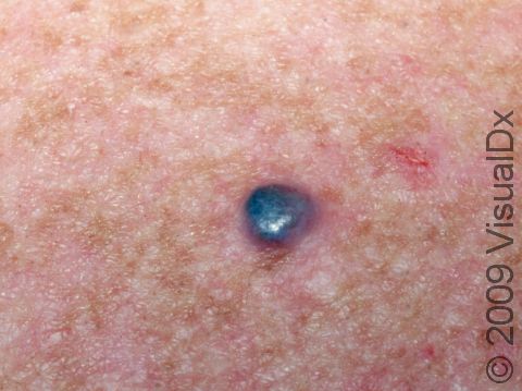
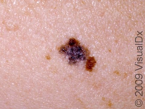
This melanoma has a classic blue-black color.
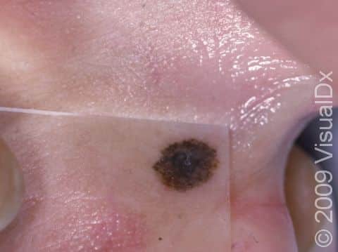
Melanoma often appears on sun-damaged skin as an irregular dark skin lesion that changes with time.
Black, multi-colored, asymmetric, or irregularly shaped lesions all need to be checked by a dermatologist or doctor skilled in looking at moles.
Graphic content
Please click to view.

Melanoma in the genital region is easily overlooked; this typical lesion on the vulva has an irregular border and both brown and blue-black colors.
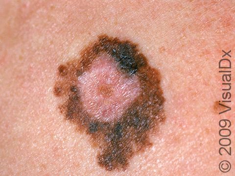
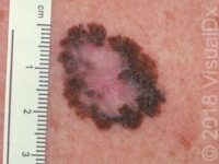
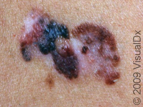
This image displays a multi-colored (including black) lesion with an irregular shape and scalloped borders typical of melanoma.
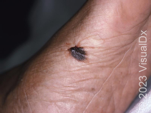
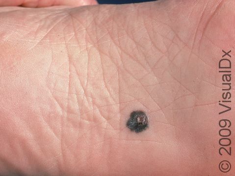
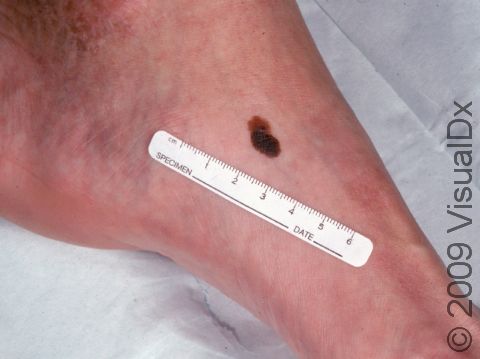
An irregularly pigmented skin lesion of the foot, consistent with malignant melanoma.
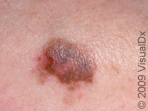
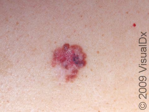
This image displays a brown, blue-gray, and pink lesion with an irregular border typical of early melanoma.
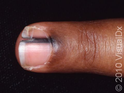
When a melanoma involves the fingernail, the cuticle often has the discoloration as well as the nail plate.
This melanoma started as a flat, irregular dark spot but has developed a raised, crusted area.
This image displays a melanoma with a white and pink center, a darker black-brown area, and pink and brown c-shaped tumor on the left side of the lesion.
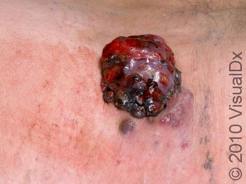
This image displays a round, bleeding melanoma that has a small "satellite" tumor underneath it.
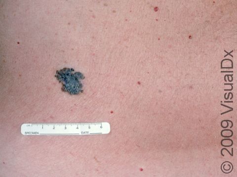
This image displays a melanoma with irregular borders surrounded by many other benign growths, which are much smaller and have regular, circular borders.
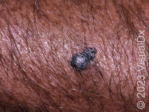
This image displays a darkly pigmented lesion typical of melanoma.
This image displays an almost black melanoma found in between the toes.
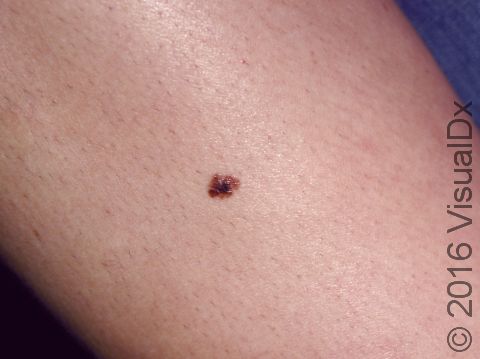
This melanoma has multiple dark colors, an asymmetrical shape, and a very irregular border typical of melanoma.
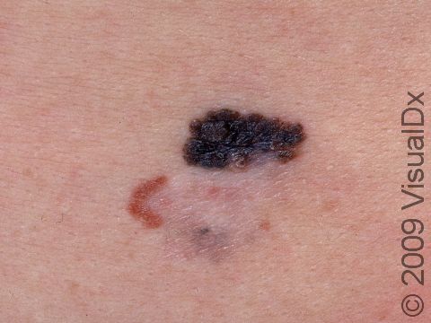
This image displays a lesion with an irregular edge and multiple colors--white, pink, pink-brown, and flecks of blue-black--typical of melanoma.
Graphic content
Melanoma
While melanoma is the least common of all the skin cancers, it is the most dangerous type of skin cancer. It can be life-threatening if it spreads (metastasizes) to other parts of the body. The frequency of diagnosis of melanoma has been increasing in recent years, faster than any other cancer. The American Cancer Society estimates that in the United States in 2023, about 97 610 new melanomas will be diagnosed, and about 7990 people are expected to die of melanoma (approximately 5420 men and 2570 women). Worldwide, an estimated 324 635 people were diagnosed with melanoma in 2020.
Melanoma starts in the pigment-producing cells of the skin (melanocytes) and may develop from an existing mole or a new mole. Early diagnosis and treatment typically lead to a complete cure, while advanced forms of melanoma can require extensive treatment. Advanced melanoma can spread to lymph nodes as well as other areas in the body, typically the lungs, liver, and brain.
Other common types of skin cancer include basal cell carcinoma and squamous cell carcinoma.
Who's At Risk?
You have an increased risk of developing melanoma if you have:
- A family history of melanoma; having someone in your family with melanoma increases your risk tenfold.
- Fair skin, light eyes, light hair, and a tendency to freckle, for example those with red or blond hair and blue or green eyes.
- A large number of moles, especially unusual-appearing moles.
- A history of frequent sun exposure.
- A history of sunburns, especially in childhood.
- A decreased immune system, such as transplant patients and individuals with HIV / AIDS.
Melanoma may be seen at any age, but it is most often diagnosed during middle age.
Using sunlamps and tanning beds can increase your risk of melanoma, especially if they cause sunburn.
Signs & Symptoms
Melanoma usually occurs on areas of the skin that are exposed to the sun, but it may be found anywhere on the body, including the eye, mouth, and genital areas.
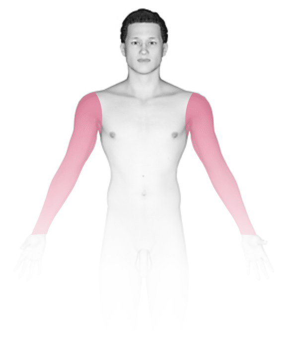
- Men are most likely to develop melanoma on the head, neck, and trunk.
- Women are most likely to develop melanoma on the legs.
- In darker skin colors, melanoma most often develops on the hands, feet, and under the nails.
A helpful tool to help you identify a growth that may be melanoma is the ABCDE checklist:
- A – Asymmetry: One half of the mole does not look like the other half.
- B – Border: The outline of the mole is irregular.
- C – Color: More than one color can be seen, such as brown, black, red, blue, or white.
- D – Diameter: A mole larger than 6 mm (1/4 inch), which is roughly the size of a pencil eraser.
- E – Evolving: Changes in the mole over time.
Self-Care Guidelines
Protective measures, such as avoiding skin exposure to sunlight during peak sun hours (10 AM to 3 PM), wearing protective clothing, and applying high sun protection factor (SPF) sunscreen, are essential for reducing exposure to harmful ultraviolet (UV) light.
Once a month, you should perform a self-exam to look for signs of skin cancer. It is best to perform the exam in a well-lit area after a shower or bath. Use a full-length mirror with the added assistance of a hand mirror when necessary. Using a hair dryer can help you examine any areas of skin covered by hair, such as your scalp.
- In front of a full-length mirror, inspect the front of your body, making sure to look at the front of your neck, chest (including under breasts), legs, and genital areas.
- With your arms raised, inspect both sides of your body, making sure to examine your underarms.
- With your elbows bent, examine the front and back of your arms as well as your elbows, hands, fingers, the areas between your fingers, and your fingernails.
- Inspect the tops and bottoms of your feet, the areas between your toes, and your toenails.
- With your back to the mirror and holding a hand mirror, inspect the back of your body, including the back of your neck, shoulders, legs, and buttocks.
- Using a hand mirror, examine your scalp and face.
As you perform your monthly self-exam, familiarize yourself with the moles, freckles, and other marks on your body, and look for any changes in them, including changes in shape, size, color, and other changes, such as moles that bleed or itch.
Treatments
A skin biopsy is needed to confirm the diagnosis, usually performed by a dermatologist.
The procedure involves:
- Numbing the skin with an injectable anesthetic.
- Sampling a small piece of skin.
- Having the skin sample examined under the microscope by a dermatopathologist.
If the diagnosis of melanoma is confirmed by biopsy, a surgical excision of the area with a margin of normal skin will need to be performed. The dermatologist may also recommend sampling of the nearby lymph nodes to see if they are involved (called a sentinel lymph node biopsy). Imaging studies of other areas of the body, such as the chest and abdomen, might be recommended if the melanoma is thick. For more advanced melanomas, an oncologist may be consulted to advise on further therapies, such as immunotherapy.
If you have previously been diagnosed and treated for melanoma, you are at increased risk of developing another melanoma, especially in the first 3 years after diagnosis. Therefore, it is essential that you regularly follow up with your medical professional to have a thorough skin examination at more frequent intervals.
Visit Urgency
If you have developed a new lesion on sun-exposed skin or if you have a spot that bleeds easily or does not seem to be healing, then you should make an appointment with a medical professional, such as a dermatologist. You should also make an appointment if an existing mole on the skin changes size, shape, color, or texture, or if it starts to itch, bleed, or become tender.
Use the ABCDE checklist described above to help you decide which existing moles are concerning for melanoma. If you have a mole that you think fits one or more of these descriptions, call a medical professional, such as a dermatologist, for evaluation.
References
American Society of Clinical Oncology. Melanoma: statistics. ASCO. https://www.cancer.net/cancer-types/melanoma/statistics. Updated 2023 Mar.
American Cancer Society. Key statistics for melanoma skin cancer. ACS. https://www.cancer.org/cancer/types/melanoma-skin-cancer/about/key-statistics.html. Updated 2023 Jan 12.
Bolognia J, Schaffer JV, Cerroni L. Dermatology. 4th ed. Philadelphia, PA: Elsevier; 2018.
James WD, Elston D, Treat JR, Rosenbach MA. Andrew’s Diseases of the Skin. 13th ed. Philadelphia, PA: Elsevier; 2019.
Kang S, Amagai M, Bruckner AL, et al. Fitzpatrick’s Dermatology. 9th ed. New York, NY: McGraw-Hill Education; 2019.
Disease Groups:
Skin Cancer and Moles
Last modified on June 26th, 2026 at 12:14 pm
Related Articles
Not sure what to look for?
Try our new Rash and Skin Condition Finder