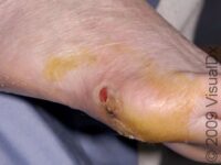
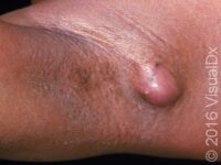
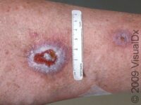
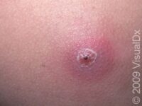
Abscess Due to an injury to the foot, an abscess formed and was then drained by the emergency doctor.
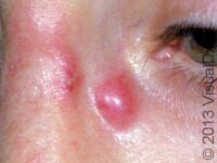
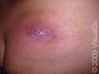
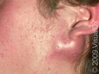
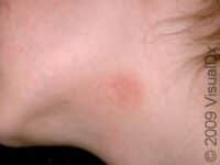
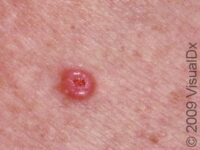
Abscess This is a typical abscess, which develops as a painful red or violaceous lump. Pushing on this with a finger or…
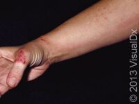
Abscess This image displays a confirmed CA-MRSA (community-associated methicillin-resistant Staphylococcal…
Abscess Due to an injury to the foot, an abscess formed and was then drained by the emergency doctor.
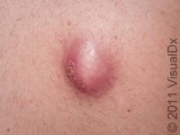
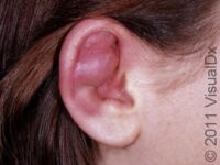
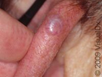
Abscess Abscesses can occur at the site of an injury. In this case, an abscess developed after ear cartilage piercing.…
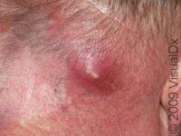
Abscess By pushing on this warm, tender abscess with a finger, there is a sense of fluid (pus) that can be felt within the…
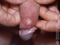
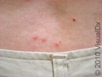
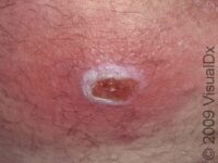
Graphic content Abscess The abscess on the shaft of the penis is red, swollen, and tender. The upper edge has a small area of broken skin…
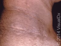
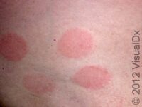
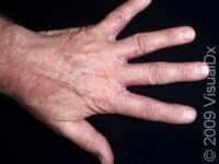
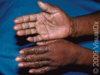
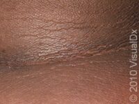
Acanthosis Nigricans This image displays acanthosis nigricans, which affects the body folds, most frequently the neck and armpits.
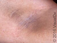
Acanthosis Nigricans Acanthosis nigricans, most commonly, is noticed at the armpits and/or neck as a slightly thickened color…
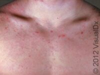
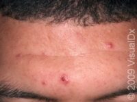
Acne (Acne Vulgaris) This image displays pus-filled lesions with whiteheads and blackheads (closed and open comedones) in an adult…
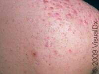
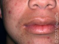
Acne (Acne Vulgaris) This image displays bumps, pus-filled lesions, whiteheads (closed comedones), and flat, brown marks from old…
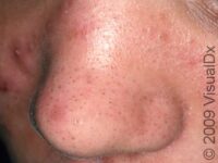
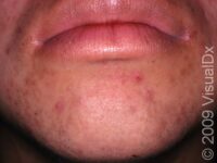
Acne (Acne Vulgaris) Multiple "blackheads" (open comedones) as well as a few red, inflammatory bumps are seen here on the nose.
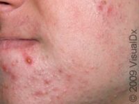
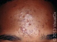
Acne (Acne Vulgaris) This image displays a mix of blackheads (open comedones), red bumps, and depressed scars typical of acne…
Acne (Acne Vulgaris) This image displays pus-filled lesions with whiteheads and blackheads (closed and open comedones) in an adult…
Acne (Acne Vulgaris) This image displays pus-filled lesions with whiteheads and blackheads (closed and open comedones) in an adult…
Acne Keloidalis Nuchae Numerous smooth, scar-like, small, raised lesions at the back of the neck are typical of acne keloidalis nuchae.
Acne Keloidalis Nuchae This image displays the back of the neck at the hairline that is affected by acne keloidalis nuchae.
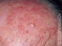
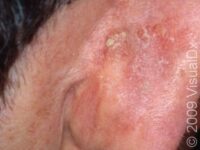
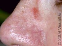
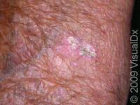
Actinic Keratosis (Solar Keratosis) This image displays an actinic keratosis in the center as well as prominent blood vessels, which suggest chronic…
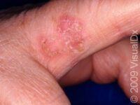
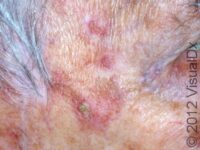
Actinic Keratosis (Solar Keratosis) This image displays an actinic keratosis in an unusual location, the side of the finger.
Actinic Keratosis (Solar Keratosis) This image displays an actinic keratosis in an unusual location, the side of the finger.
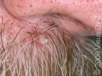
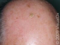
Actinic Keratosis (Solar Keratosis) This image displays a bald scalp with areas of sun damage and actinic keratoses.
Actinic Keratosis (Solar Keratosis) The surface of the ear is a typical location of actinic keratoses.
Actinic Keratosis (Solar Keratosis) Actinic keratoses are pre-cancerous, due to too much sun-exposure over years, which appear as small, scaly…
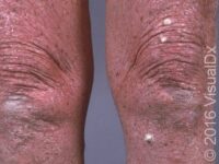
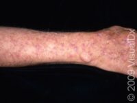
Actinic Keratosis (Solar Keratosis) The forearm is a very common area for sun damage and actinic keratoses.
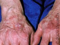
Actinic Keratosis (Solar Keratosis) This image displays numerous areas of sun damage and actinic keratoses.
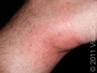
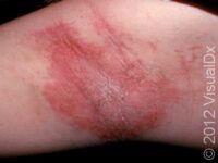
Allergic Contact Dermatitis Contact dermatitis often has slightly elevated lesions with distinct borders.
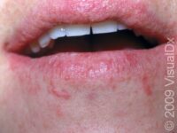
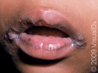
Allergic Contact Dermatitis This image displays redness around the mouth caused by an allergic reaction to mangoes.
Allergic Contact Dermatitis This image displays redness around the mouth caused by an allergic reaction to mangoes.
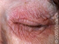
Allergic Contact Dermatitis The thin eyelid skin is a frequent site for allergic contact dermatitis due to inadvertent touching the eyelids…
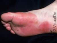
Allergic Contact Dermatitis The sharp border of the redness on the foot is due to contact dermatitis from an allergy to a substance in contact…
Allergic Contact Dermatitis This hairdresser had an allergic contact dermatitis from exposure to hair dye.
Allergic Contact Dermatitis This image displays a scaly, slightly elevated lesion due to an allergy to the nickel in an eyeglass frame.
Allergic Contact Dermatitis This image displays contact dermatitis, also called "fiddler's neck," from an allergy to the violin touching…
Allergic Contact Dermatitis This image displays redness around the mouth caused by an allergic reaction to mangoes.
Graphic content Allergic Contact Dermatitis This image displays a violet-colored, linear, slightly elevated lesion typical of contact dermatitis, due to…
Allergic Contact Dermatitis This hairdresser had an allergic contact dermatitis from exposure to hair dye.
Allergic Contact Dermatitis This hairdresser had an allergic contact dermatitis from exposure to hair dye.
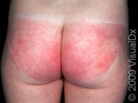
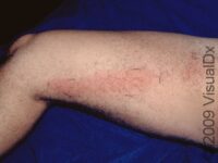
Allergic Contact Dermatitis Contact dermatitis that has been present for longer periods of time can appear like many other rashes, with…
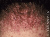
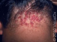
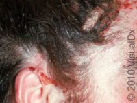
Allergic Contact Dermatitis This image displays contact dermatitis on the scalp and adjacent to the scalp area in a young man who was using a…
Allergic Contact Dermatitis This image displays contact dermatitis on the scalp and adjacent to the scalp area in a young man who was using a…
Allergic Contact Dermatitis This is severe allergic contact dermatitis, resulting in very thick, scaly lesions on the fingers.
Allergic Contact Dermatitis This is severe allergic contact dermatitis, resulting in very thick, scaly lesions on the fingers.
Allergic Contact Dermatitis This image displays allergic contact dermatitis from fragrance found in a deodorant.
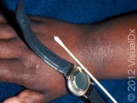
Allergic Contact Dermatitis This image displays an allergy to the nickel found in the watch case. The result is a scaly, itchy, persistent skin…
Allergic Contact Dermatitis Contact dermatitis often has slightly elevated lesions with distinct borders.
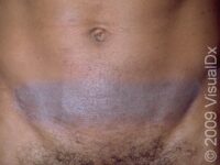
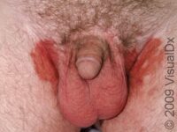
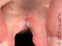
Graphic content Allergic Contact Dermatitis The scrotum and penis are frequent sites of contact dermatitis.
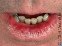
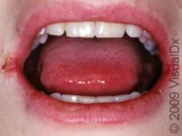
Angular Cheilitis This image displays a frequent location for candida infection (angular cheilitis), the corners of the mouth.
Angular Cheilitis The cracking at the corners of the mouth in oral candidiasis, as displayed in this image, is known as angular…
Angular Cheilitis This image displays a frequent location for candida infection (angular cheilitis), the corners of the mouth.
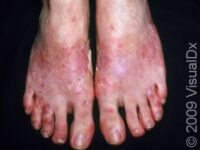
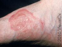
Athlete’s Foot (Tinea Pedis) This image displays a scaly border and pink, slightly elevated lesions typical of tinea pedis (athlete's foot…
Athlete’s Foot (Tinea Pedis) This image displays a scaly border and pink, slightly elevated lesions typical of tinea pedis (athlete's foot…
Athlete’s Foot (Tinea Pedis) This image displays a scaly border and pink, slightly elevated lesions typical of tinea pedis (athlete's foot…
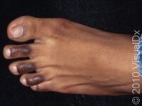
Athlete’s Foot (Tinea Pedis) This image displays the fungal infection that frequently occurs between the toes, tinea pedis (athlete's…
Athlete’s Foot (Tinea Pedis) This image displays the fungal infection that frequently occurs between the toes, tinea pedis (athlete's…
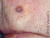
Basal Cell Carcinoma (BCC) Basal cell carcinomas may sometimes have a blue-black, irregular discoloration.
Graphic content Basal Cell Carcinoma (BCC) Basal cell carcinomas can grow rapidly, in weeks to months, or slowly, over years.
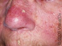
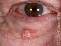
Basal Cell Carcinoma (BCC) The nodular form of basal cell carcinoma is usually skin-colored with tiny blood vessels visible.
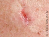
Basal Cell Carcinoma (BCC) The infiltrating type of basal cell carcinoma can appear as a scar or resemble a superficial skin ulcer. These…
Basal Cell Carcinoma (BCC) The nodular form of basal cell carcinoma is usually skin-colored with tiny blood vessels visible.
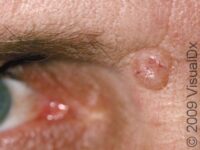
Basal Cell Carcinoma (BCC) This image displays a shiny-appearing lesion with small, visible blood vessels typical of basal cell…