Actinic keratoses are precancerous, due to too much sun exposure over years, which appear as small, scaly lesions that are rough to the touch.
This image displays actinic keratosis of the legs.
The surface of the ear is a typical location of actinic keratoses.
This image displays a bald scalp with areas of sun damage and actinic keratoses.
This image displays actinic keratoses that have developed very thick, crusty scale known as hypertrophic actinic keratoses.
This image displays a frequent site for actinic keratoses, the rim of the ear.
This image displays actinic keratoses hidden by facial hair.
This image displays an actinic keratosis in the center as well as prominent blood vessels, which suggest chronic sun damage.
This image displays a close-up of actinic keratoses.
This image displays an actinic keratosis with brown spots suggesting chronic sun damage.
This image displays a thin white scale typical of actinic keratosis.
This image displays multiple actinic keratoses on the face.
This image displays numerous areas of sun damage and actinic keratoses.
This image displays a rather large, inflamed actinic keratosis.
This image displays a very large, scaly actinic keratosis.
This image displays two actinic keratoses and thinning and wrinkling of the skin, which suggests severe, chronic sun damage.
The forearm is a very common area for sun damage and actinic keratoses.
This image displays a large actinic keratosis with a yellowish crust.
This image displays an actinic keratosis in an unusual location, the side of the finger.
While the rough, gritty scale of actinic keratoses is sometimes easier to feel than see, it can also be very obvious to the naked eye as with this lesion on the forehead.

Images of Actinic Keratosis (Solar Keratosis) (20)

Actinic keratoses are precancerous, due to too much sun exposure over years, which appear as small, scaly lesions that are rough to the touch.
This image displays actinic keratosis of the legs.
The surface of the ear is a typical location of actinic keratoses.
This image displays a bald scalp with areas of sun damage and actinic keratoses.
This image displays actinic keratoses that have developed very thick, crusty scale known as hypertrophic actinic keratoses.
This image displays a frequent site for actinic keratoses, the rim of the ear.
This image displays actinic keratoses hidden by facial hair.
This image displays an actinic keratosis in the center as well as prominent blood vessels, which suggest chronic sun damage.
This image displays a close-up of actinic keratoses.
This image displays an actinic keratosis with brown spots suggesting chronic sun damage.
This image displays a thin white scale typical of actinic keratosis.
This image displays multiple actinic keratoses on the face.
This image displays numerous areas of sun damage and actinic keratoses.
This image displays a rather large, inflamed actinic keratosis.
This image displays a very large, scaly actinic keratosis.
This image displays two actinic keratoses and thinning and wrinkling of the skin, which suggests severe, chronic sun damage.
The forearm is a very common area for sun damage and actinic keratoses.
This image displays a large actinic keratosis with a yellowish crust.
This image displays an actinic keratosis in an unusual location, the side of the finger.
While the rough, gritty scale of actinic keratoses is sometimes easier to feel than see, it can also be very obvious to the naked eye as with this lesion on the forehead.

Actinic Keratosis (Solar Keratosis)

Actinic keratoses, also known as solar keratoses, are small rough, scaly, slightly raised areas of skin (papules) that usually occur in body locations that have been chronically exposed to the sun. Actinic keratoses are precancerous, each having a less than 1% chance of turning into a skin cancer (squamous cell skin cancer). Because of this risk, it is important to perform self-examinations regularly and get them treated by a medical professional early.

Who's At Risk?

People most at risk for actinic keratosis are those with the lightest of skin colors who sunburn easily, as well as people with blue, green, or hazel eyes and red or blond hair. People with light skin colors who have had a lot of sun exposure over many years are at a high risk, as well. Those with a weak immune system due to chemotherapy, HIV, or an organ transplant are also at higher risk.

Actinic keratoses usually appear in those who are older than 50, but they can start appearing in younger adults who have had a lot of sun exposure. Those with darker skin colors can be affected but at lower rates.

Signs & Symptoms

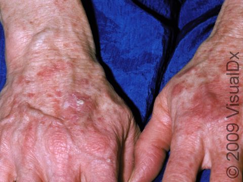
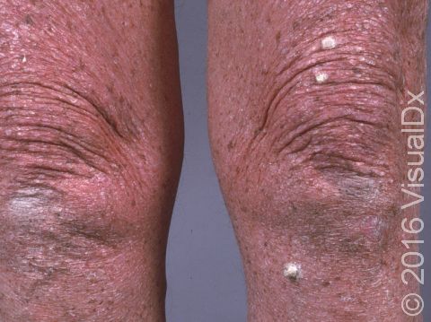
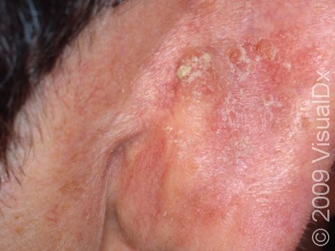
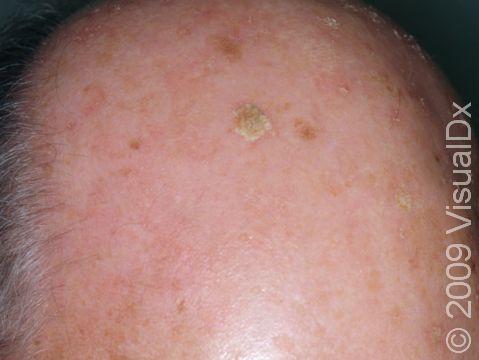
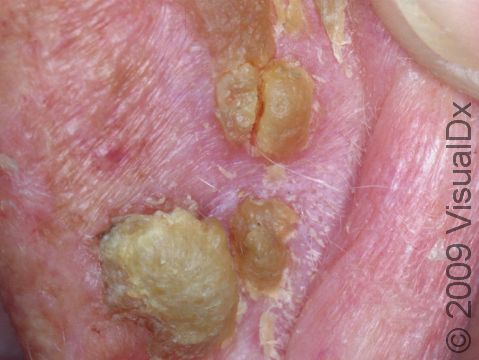
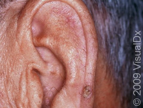
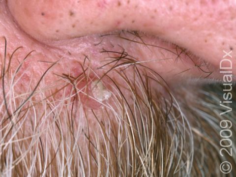
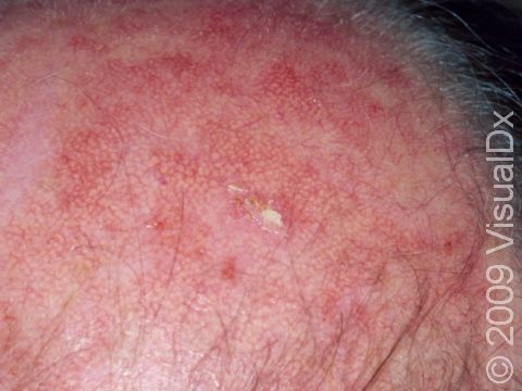
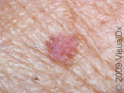
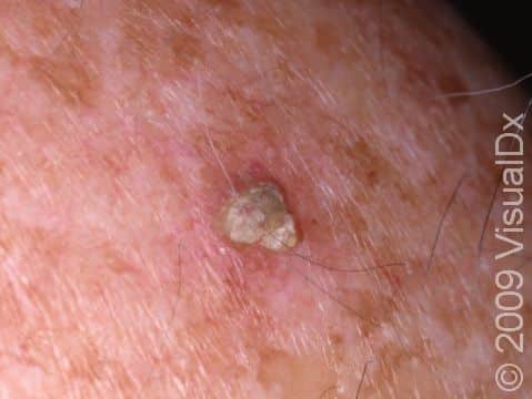
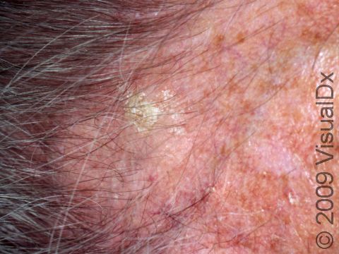
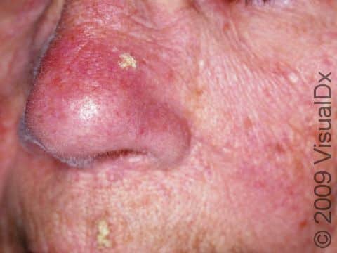
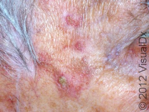
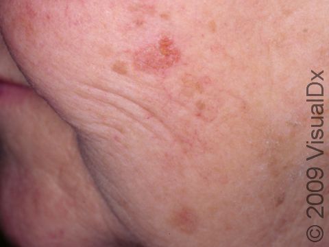
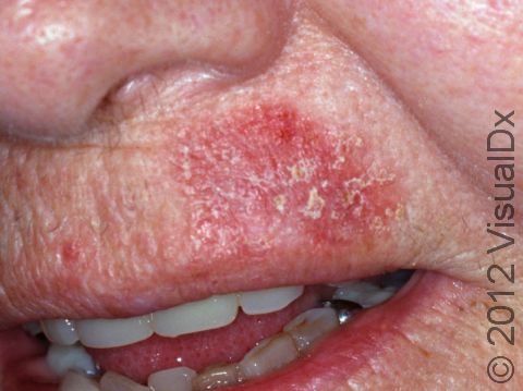
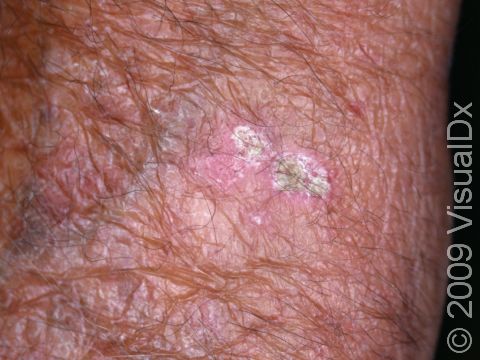
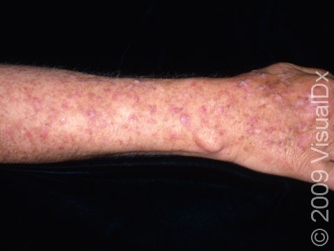
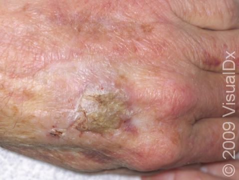
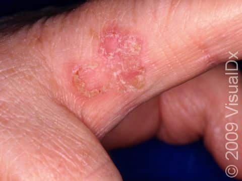
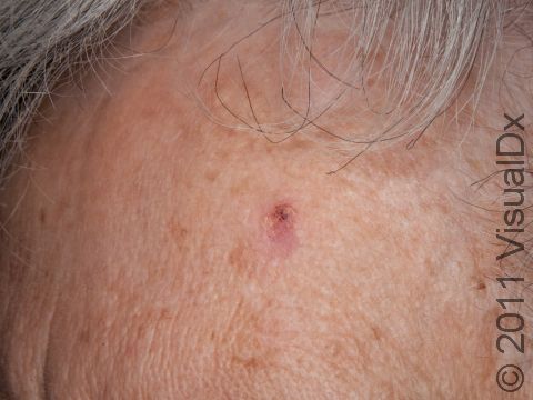
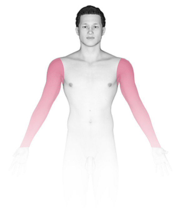
Actinic keratoses are rough, scaly, slightly raised papules on sun-damaged skin. The face, scalp (if balding), ears, neck, forearms, and backs of the hands are most commonly affected with actinic keratoses, but any skin area frequently exposed to sun can be affected.

Actinic keratoses vary in size. They have slight scale (sometimes thick like a wart) and a pink, red, or brownish color. They are slightly rough, like fine sandpaper, and may be a bit sensitive to the touch.

Self-Care Guidelines

Prevention is very important. Sun protection can reduce the number of new actinic keratoses from occurring and may help small lesions go away on their own.

  • Avoid direct sun in the middle of the day (10 AM to 3 PM). Be mindful that snow and water reflect light to the skin. Also note that clouds still let a lot of light through, so you can be exposed to ultraviolet light even on cloudy days.
  • Wear a hat with a wide brim. A baseball hat does not give as much protection.
  • Cover up with tightly woven clothing. Some manufacturers make specialty clothing with an ultraviolet protection factor (UPF) rating.
  • Use sunscreen on all exposed skin areas, including the lips, before going outdoors. A broad-spectrum sunscreen (meaning it blocks UVB and UVA light) with an SPF of at least 30 is best. Apply sunscreen generously 30 minutes before going outdoors, and reapply every 2 hours or after swimming or sweating a lot. Sunscreen sprays tend not to provide a thick enough layer on your skin. Therefore, sunscreen lotions or creams are recommended.
  • Avoid using tanning beds.

Perform a self-exam regularly to look for signs of skin cancer. It is best to perform the exam in a well-lit area following a shower or bath. Use a full-length mirror with the added assistance of a hand mirror, as necessary. Using a hair dryer can help you examine any areas of skin covered by hair, such as your scalp.

  • In front of a full-length mirror, inspect the front of your body, making sure to look at your neck, chest (including under breasts), legs, and genitals.
  • With your arms raised, inspect both sides of your body, making sure to examine your underarms.
  • With your elbows bent, examine the front and back of your arms as well as your elbows, hands, fingers, area between your fingers, and your fingernails.
  • Inspect the tops and bottoms of your feet, the area between your toes, and your toenails.
  • With your back to the mirror and holding a hand mirror, inspect the back of your neck, shoulders, legs, and buttocks.
  • Using a hand mirror, examine your scalp and face.

As you perform your self-exam, familiarize yourself with the moles, freckles, and other marks on your body, and look for any changes in them from month to month, including changes in shape, size, color, and other changes, such as moles that bleed or itch.

Treatments

Depending on how many actinic keratoses you have, your medical professional will tailor treatment, which may include:

  • Removal with freezing (cryosurgery), scraping (curettage), or burning (electrocautery).
  • Creams with either fluorouracil (eg, Carac), fluorouracil and calcipotriene, imiquimod (eg, Zyclara), diclofenac gel, or tirbanibulin (Klisyri).
  • Chemical peeling.
  • Photodynamic therapy.

Visit Urgency

If you have a new skin growth or an existing lesion that is growing or bleeding, see your medical professional.

References

Bolognia J, Schaffer JV, Cerroni L. Dermatology. 4th ed. Philadelphia, PA: Elsevier; 2018.

James WD, Elston D, Treat JR, Rosenbach MA. Andrew’s Diseases of the Skin. 13th ed. Philadelphia, PA: Elsevier; 2019.

Kang S, Amagai M, Bruckner AL, et al. Fitzpatrick’s Dermatology. 9th ed. New York, NY: McGraw-Hill Education; 2019.

Last modified on May 30th, 2023 at 2:07 pm

Not sure what to look for?

Try our new Rash and Skin Condition Finder

Age
Gender
Submit