The thickened irregular surface of a seborrheic keratosis of the skin.
This image displays multiple brown, slightly elevated lesions typical of seborrheic keratoses.
Seborrheic keratoses are common, benign skin lesions in adults. They have a
Seborrheic keratoses are common, benign skin lesions in adults. They have a "stuck on" and rough surface appearance.
Widespread seborrheic keratoses of the trunk in an individual with darkly pigmented skin.
Seborrheic keratoses have a
Seborrheic keratoses have a "stuck on" or rough surface appearance, which can be skin-colored or dark.
A waxy, tan-colored,
A waxy, tan-colored, "stuck-on-appearing" lesion is characteristic of a seborrheic keratosis.

Graphic content

Please click to view.

This image displays the waxy and stuck-on appearing, elevated lesion typical of a seborrheic keratosis.
Seborrheic keratoses are harmless thickenings of the outer layer of skin.
Pigmented seborrheic keratosis of the skin with its characteristic warty-like texture.
This image displays a raised lesion typical of seborrheic keratosis.
A seborrheic keratosis lesion can sometimes become black, as displayed in this image.
This image displays a lesion, with a sharp border and rough surface that appears to be sitting on top of the skin, typical of seborrheic keratosis.
Dark brown, rough seborrheic keratoses may be found on any skin area, even inside the ear rim.
Seborrheic keratoses may become very large and lumpy in the scalp area. There are several on this man, which have grown into each other over time.
Typical to seborrheic keratoses, this image displays a brown, rough-appearing lesion.
The dark to light brown raised, rough areas of seborrheic keratoses may be numerous in the elderly.
The multiple small, light brown freckles seen here reflect sun damage, but the darker, larger spot on the upper nose bridge is a rough, scaling seborrheic keratosis.

Images of Seborrheic Keratosis: Symptoms and Treatment (18)

The thickened irregular surface of a seborrheic keratosis of the skin.
This image displays multiple brown, slightly elevated lesions typical of seborrheic keratoses.
Seborrheic keratoses are common, benign skin lesions in adults. They have a
Widespread seborrheic keratoses of the trunk in an individual with darkly pigmented skin.
Seborrheic keratoses have a
A waxy, tan-colored,

Graphic content

This image displays the waxy and stuck-on appearing, elevated lesion typical of a seborrheic keratosis.
Seborrheic keratoses are harmless thickenings of the outer layer of skin.
Pigmented seborrheic keratosis of the skin with its characteristic warty-like texture.
This image displays a raised lesion typical of seborrheic keratosis.
A seborrheic keratosis lesion can sometimes become black, as displayed in this image.
This image displays a lesion, with a sharp border and rough surface that appears to be sitting on top of the skin, typical of seborrheic keratosis.
Dark brown, rough seborrheic keratoses may be found on any skin area, even inside the ear rim.
Seborrheic keratoses may become very large and lumpy in the scalp area. There are several on this man, which have grown into each other over time.
Typical to seborrheic keratoses, this image displays a brown, rough-appearing lesion.
The dark to light brown raised, rough areas of seborrheic keratoses may be numerous in the elderly.
The multiple small, light brown freckles seen here reflect sun damage, but the darker, larger spot on the upper nose bridge is a rough, scaling seborrheic keratosis.

Seborrheic Keratosis: Symptoms and Treatment

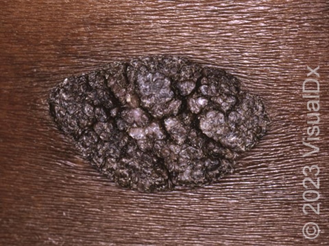
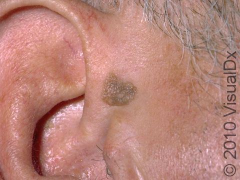
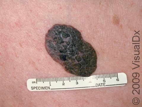
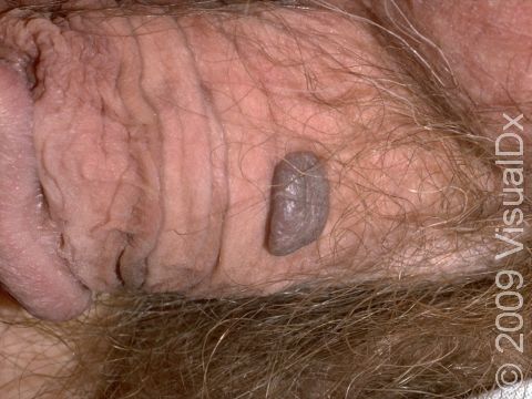
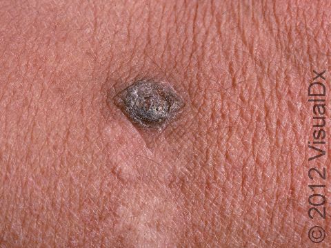
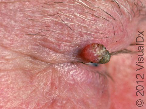
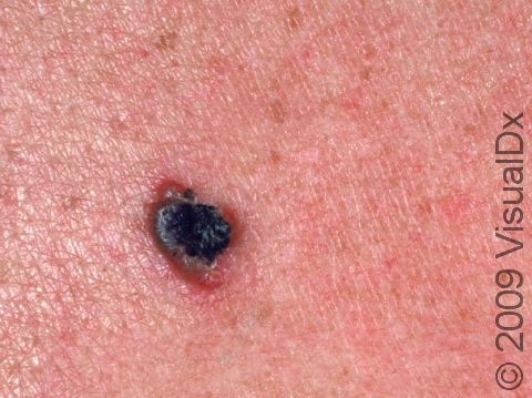
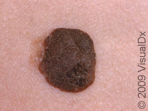
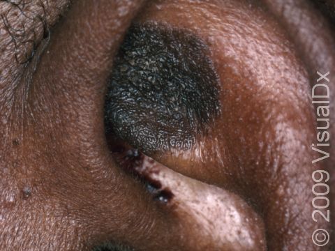
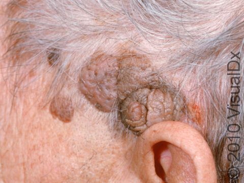
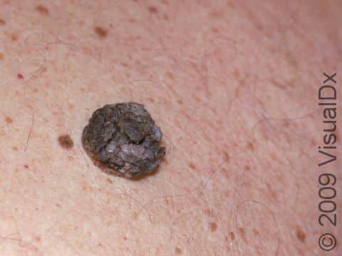
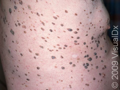
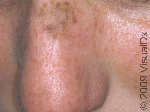
Seborrheic keratoses are common noncancerous (benign) growths of unknown cause seen in adults due to a thickening of an area of the top skin layer. Seborrheic keratoses may appear as if they are stuck on to the skin. They have distinct borders, and they may appear as papules (small, solid bumps) or plaques (solid, raised patches that are bigger than a thumbnail). They may be the same color as your skin, or they may be pink, light brown, darker brown, or very dark brown, or sometimes may appear black.

Who's At Risk?

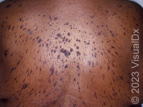
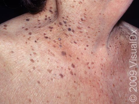
Seborrheic keratoses can occur any time after puberty, and almost everyone older than 50 has one or more of these skin growths. They may increase in number with age. Members of the same family can have an inherited tendency to grow multiple seborrheic keratoses. Men and women are equally as likely to develop them. People with darker skin colors tend to develop seborrheic keratoses less frequently than those with lighter skin colors.

Signs & Symptoms

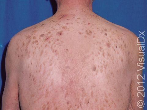
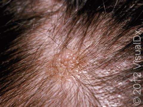
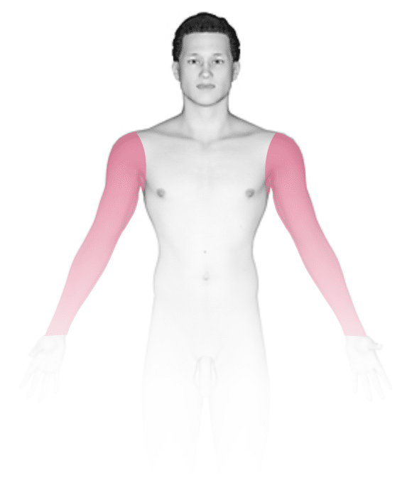
Seborrheic keratoses can occur anywhere on the body, except for the palms, soles, and mucous membranes (areas such as in the mouth or anus). They most commonly occur on the chest and back. Seborrheic keratoses do not go away on their own, and they do not become cancerous.

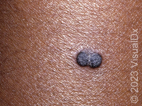
They usually start as light brown or skin-colored, slightly raised areas, which can be round or oval and of varying size (usually smaller than a thumbnail, but sometimes much larger). As they grow thicker, seborrheic keratoses may become dark brown to almost black and appear to be “stuck on” to the surface of the skin. The surface may feel smooth or rough. In lighter skin colors, they may be pink or any shade of brown. In darker skin colors, they may be any shade of brown, purple, gray, or blackish.

Self-Care Guidelines

No treatment is needed unless there is irritation from clothing, such as itching or bleeding.

Note that:

  • There is no way to prevent new seborrheic keratoses from forming.
  • Some lotions containing alpha hydroxy acids, salicylic acid, or urea may make the areas feel smoother with regular use but will not eliminate them.
  • Over-the-counter freezing techniques are available but are usually not effective.


Treatments

Removal can be accomplished with freezing (cryosurgery), scraping (curettage), burning (electrocautery), lasers, or acids. Your dermatologist or other medical professional might conduct a biopsy if the growth looks unusual.

Visit Urgency

If a lesion on your skin is growing, bleeding, painful, or itchy, see your dermatologist or another medical professional. Similarly, consult a medical professional for any growth that is more than one color, that is dark brown or black, or that looks different than any of your other skin growths.

Seborrheic keratoses can be removed, but removal is considered a cosmetic issue and is usually not covered by insurance.

References

Bolognia J, Schaffer JV, Cerroni L. Dermatology. 4th ed. Philadelphia, PA: Elsevier; 2018.

James WD, Elston D, Treat JR, Rosenbach MA. Andrew’s Diseases of the Skin. 13th ed. Philadelphia, PA: Elsevier; 2019.

Kang S, Amagai M, Bruckner AL, et al. Fitzpatrick’s Dermatology. 9th ed. New York, NY: McGraw-Hill Education; 2019.

Last modified on September 28th, 2023 at 5:17 pm

Not sure what to look for?

Try our new Rash and Skin Condition Finder

Age
Gender
Submit