Spider Angioma
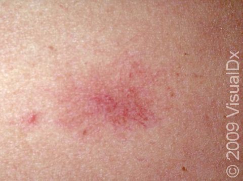
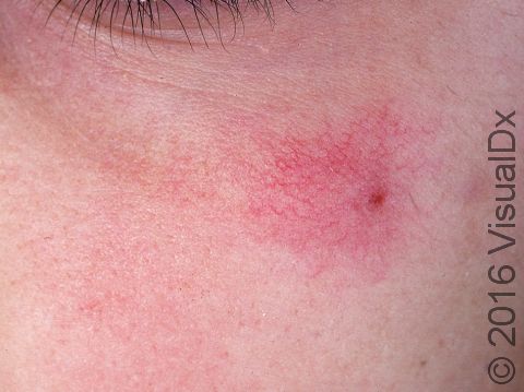
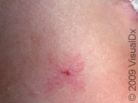
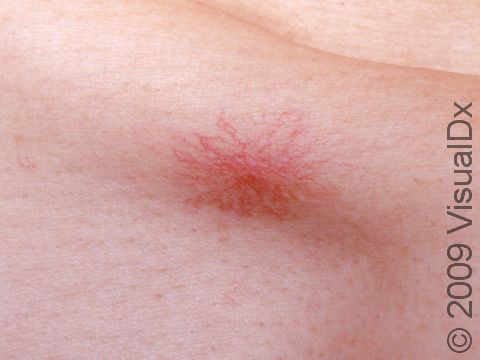
A spider angioma is a common skin condition that appears as a red papule (a small, solid bump) with small red vessels radiating from it on the surface of the skin. The pattern is thought to resemble the body and legs of a spider. Applying pressure on the central portion of a spider angioma may cause the entire lesion to disappear, but then it will rapidly refill with blood once the pressure is released.
Spider angiomas are quite common in children, but as the child grows older, the spider angioma usually fades and even disappears completely.
Who's At Risk?
Spider angiomas occur in both children and adults. Children of all races / ethnicities can develop spider angiomas, but they are more apparent in lighter skin colors. Girls and boys seem to be equally affected.
It is estimated that up to 50% of children may develop a spider angioma at some point during childhood.
Signs & Symptoms
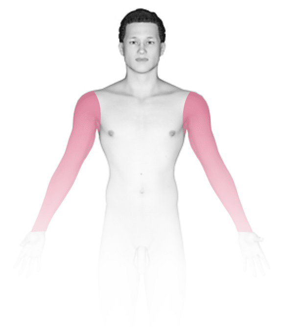
The most common locations for spider angiomas include the:
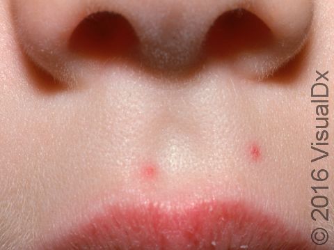
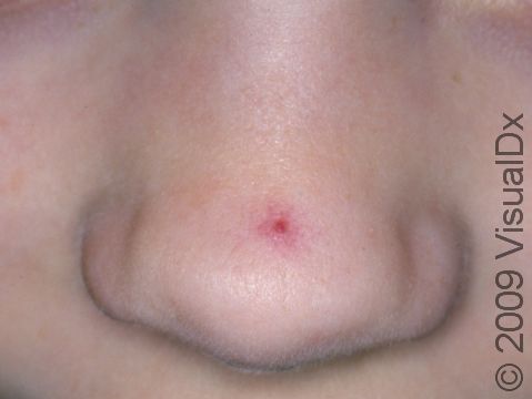
- Face, especially below the eyes and over the cheekbones.
- Neck.
- Upper trunk.
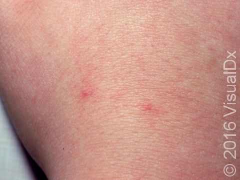
- Backs of the hands and fingers.
- Forearms.
- Ears.
Spider angiomas may appear singly or as multiple lesions. Each spider angioma appears as a small (1-10 mm), bright red spot. Upon closer inspection, you will see a central red dot with tiny red lines radiating out from the center.
Rarely, spider angiomas may bleed if injured.
Self-Care Guidelines
In children, spider angiomas usually go away without treatment, although they may take several years to disappear completely.
Treatments
No treatment is necessary for spider angiomas. However, if your child is self-conscious about the area or if it frequently bleeds, their medical professional may offer removal via:
- An electric needle (electrodesiccation).
- Laser treatment.
Visit Urgency
If the area frequently bleeds or if it begins to change in size or color, see your child’s medical professional or a dermatologist for evaluation. The child should also be evaluated if multiple angiomas suddenly develop at the same time.
References
Bolognia J, Schaffer JV, Cerroni L. Dermatology. 4th ed. Philadelphia, PA: Elsevier; 2018.
James WD, Elston D, Treat JR, Rosenbach MA. Andrew’s Diseases of the Skin. 13th ed. Philadelphia, PA: Elsevier; 2019.
Kang S, Amagai M, Bruckner AL, et al. Fitzpatrick’s Dermatology. 9th ed. New York, NY: McGraw-Hill Education; 2019.
Paller A, Mancini A. Paller and Mancini: Hurwitz Clinical Pediatric Dermatology. 6th ed. St. Louis, MO: Elsevier; 2022.
Last modified on June 26th, 2024 at 12:15 pm
Not sure what to look for?
Try our new Rash and Skin Condition Finder